Socket anatomy
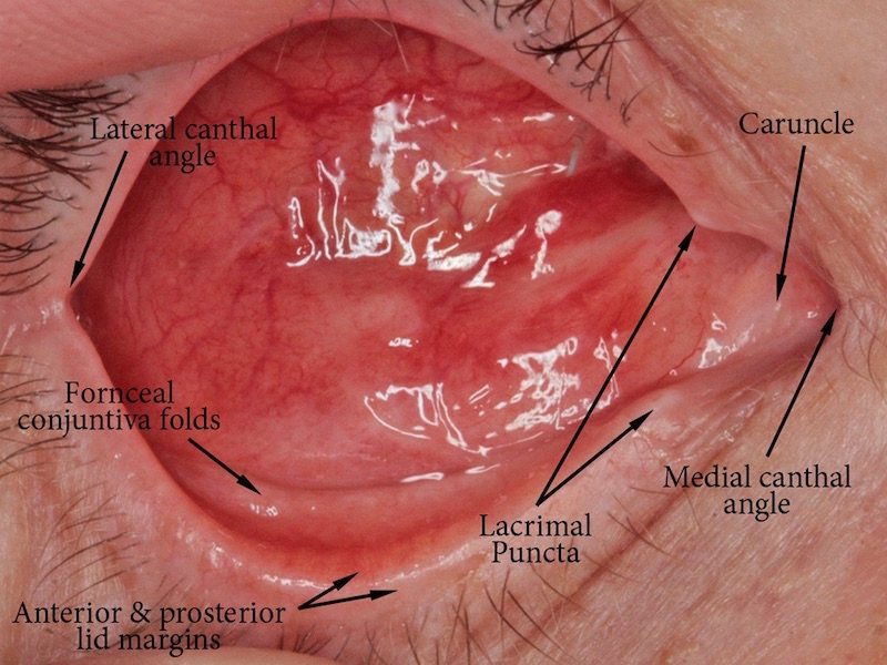

Many people (eye health professionals included) are apprehensive about seeing what is behind the eyelids of somebody who has lost an eye. The above image is designed to demonstrate that the anatomy of an anophthalmic socket is virtually identical to a normal socket – the only thing missing is the eye itself.
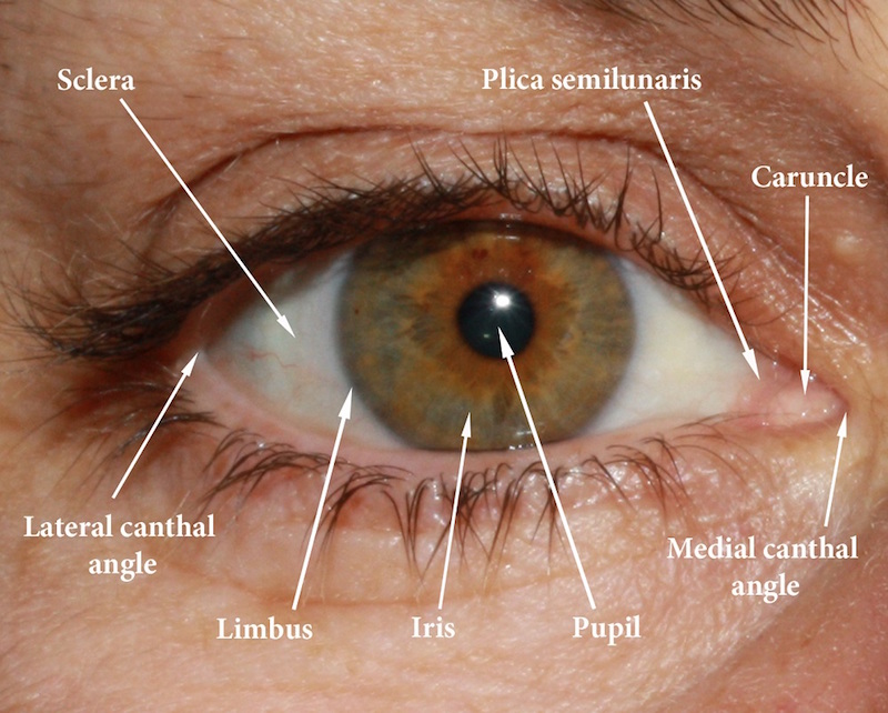
Basic anatomy of the eye.

The tear glands and ducts operate normally once a prosthetic eye is inserted in the socket.
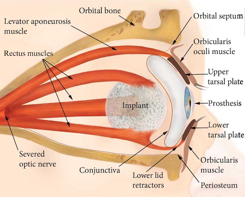
Socket anatomy with orbital implant and prosthetic eye.
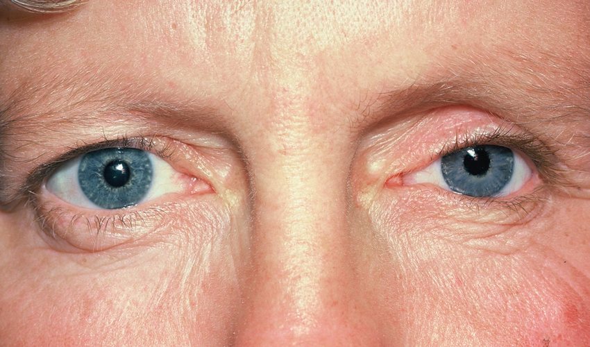
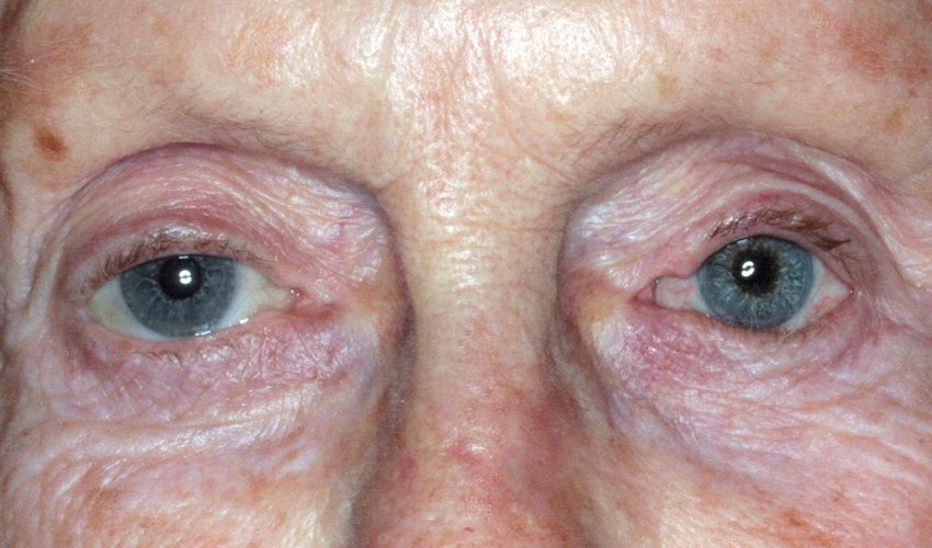
These images illustrate facial change in the same woman over a 40 year period. She was forty when the first photo was taken and recently received her latest left prosthesis at age 80.
